

")
What Pathologies Can be Visualized with Hip Ultrasound?
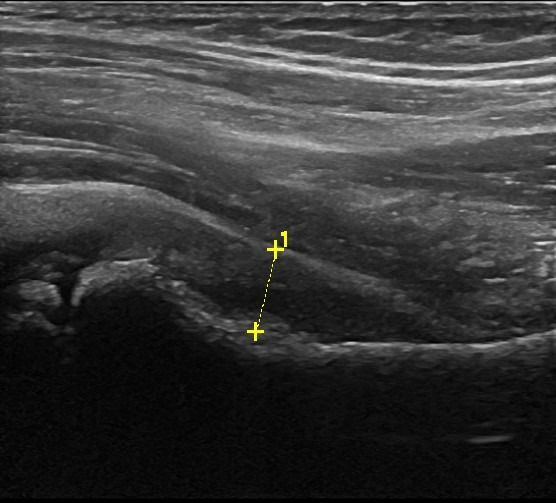
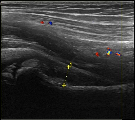
Hip Ultrasound can diagnose a range of pathologies and injury in the joint, tendons, bursae, and nerves of the hip area, such as:
Hip Joint
- Fluid Collection
- Synovitis as a symptom of Rheumatic Disease
- Osteophytes as an indication of Osteoarthritis
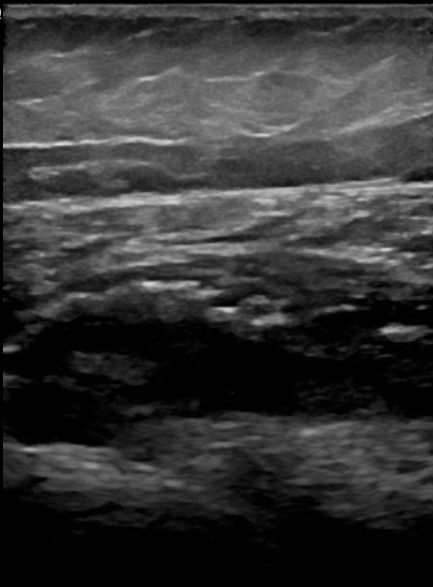
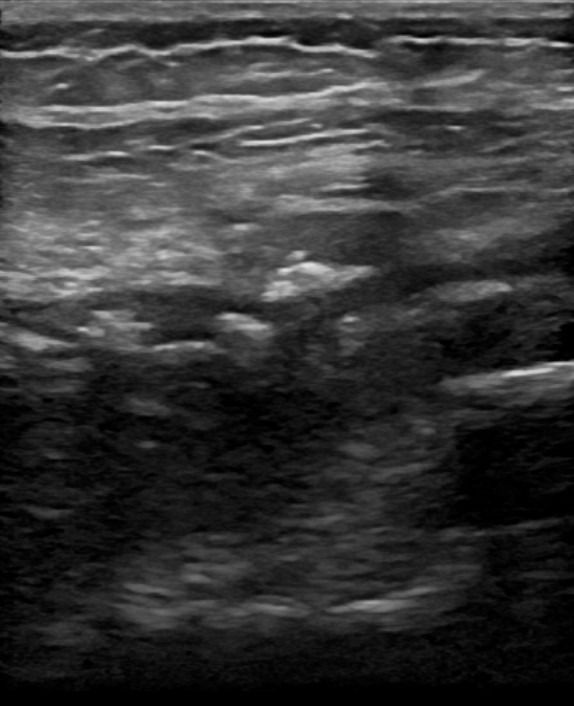
Hip Tendons and Bursae
Tendonitis or rupture of gluteal tendons, rectus femoris, and hamstrings
- Trochanteric Bursitis: fluid collection in the bursae of the gluteal tendons in the greater trochanter
- Bursitis: Fluid collection in the bursa of the tensor fasciae latae tendon
- Snapping Hip: Tendon snapping of the iliopsoas tendon or the iliotibial band
Hip Nerves
- Edema in small nerves causing Meralgia Paresthetica
- Pathologies of the Sciatic Nerve
Advantages of Hip Ultrasound Compared to Other Imaging Methods
Compared to Magnetic Resonance Imaging (MRI) and Computed Tomography (CT), Hip Ultrasound offers specific advantages:
- Dynamic Testing: Ultrasound is a dynamic examination, meaning diagnostic information can be obtained during movement. This is particularly important for hip examinations, allowing the assessment of snapping hip syndrome that may involve the internal tendon of the iliopsoas or the external iliotibial band.
- Patient-Friendly Examination: Hip Ultrasound is a safe, painless, and flexible examination suitable for all patients. It does not involve the use of intravenous drugs, exposure to radiation, or magnetic resonance coordination.
How is Hip Ultrasound Performed, and What Preparation is Needed?
The procedure of Hip Ultrasound is simple, painless, and does not require preparation from the patient. During the examination, the patient lies on the examination bed, the physician applies gel to the skin, and performs the examination with an ultrasound transducer. Under the guidance of the radiologist, the patient performs various movements with their leg as part of dynamic testing. The total duration of the examination is approximately 30 minutes.


