

")
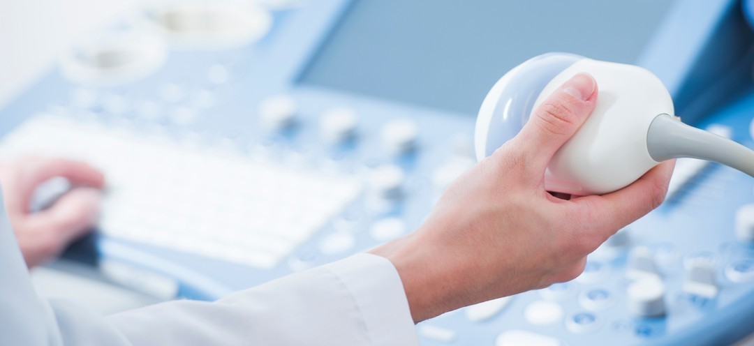 Thanks to its non-invasive nature and the extension of its applications to almost all the organs of the human body, Ultrasonography has emerged as one of the primary tools of Preventive Healthcare.
Ultrasound is a diagnostic examination that can be the first and often the only diagnostic test in the prevention of many diseases, without burdening the body with ionizing radiation or the administration of intravenous contrast agents.
As Ultrasound is a real-time examination, the specialization and experience of the Radiologist performing it, as well as the technological features of the ultrasound equipment, are of paramount importance. The ability to apply Advanced Ultrasonography Techniques, such as Elastography, Quantification Techniques, and Sensitive Vascular Detection Techniques, is crucial for the reliability of diagnosis and the effective prevention of health problems.
Ultrasound Check-Up
The organs most frequently examined with Ultrasound Check-Up are:
- Compact organs in the upper and lower abdomen: Upper Abdomen Ultrasound allows the study of the liver, gallbladder, bile ducts, pancreas, spleen, and kidneys. Lower Abdomen Ultrasound examines the bladder and internal reproductive organs, namely the ovaries, uterus, and prostate.
- Thyroid gland: The triple study of the Thyroid Gland, including B-Mode, Color Doppler Ultrasound, and Elastography, provides a comprehensive visualization of the thyroid. The outline, vascularity, elasticity of the gland, as well as and texture of a possible lesion are accurately and reliably assessed. Additionally, ultrasound enables the visualization of the parathyroid glands and the lymph nodes of the area.
- Breasts: Modern ultrasound machines allow for the analysis of breast architecture with exceptional precision. Elastography can highlight the elasticity of the lesion, indicating whether it is hard or soft. Through this technique, we can assess the nature of the lesion, providing indications of whether it is benign or malignant. Additionally, with the contribution of Color Doppler, Power Doppler, and B-Flow, we can monitor vascular flow within the lesions, offering even more information about the type and nature of the alteration.
Ultrasound Check-Up can also diagnose vascular problems such as:
- Carotid Stenosis or Occlusion: The Carotid and Vertebral Artery Triplex examines the vessels that supply blood to the brain and face. The degree of stenosis in a carotid artery is related to the likelihood of the patient experiencing a cerebrovascular event. Additionally, the quality and stability of the atherosclerotic plaque determine the chances of it detaching and causing a cerebrovascular event.
- Narrowing or Occlusion of Lower Limb Arteries: With Lower Limb Artery Triplex, pathologies such as aneurysms and stenoses can be detected. These conditions can lead to serious consequences if not identified early.
- Venous Thrombosis or Insufficiency in the Venous Network of the Lower Limbs: The Lower Limb Vein Triplex allows us to detect cases of deep or superficial vein thrombosis and examine the presence of insufficiency in the veins (varicose veins). The presence of thrombosis is a critical element for the patient's life and requires immediate intervention and pharmacological treatment.
- Diagnosis of Aneurysm or Stenosis of the Abdominal Aorta: The diagnostic information provided by the Abdominal Aorta and Iliac Vessels Triplex is particularly important. The presence of aneurysms in the abdominal aorta is a serious condition that can threaten the patient's life. Early diagnosis allows for the necessary medical measures to be taken before the pathology progresses to an urgent situation.
Personalized Approach
Within the framework of Personalized Medicine which, in recent years, has been implemented in the most advanced healthcare systems worldwide, Ultrasound Check-Up is tailored to the individual’s familial and personal medical history, while simultaneously relying on possible findings from the examination by the Clinical Doctor.
Personalized and regular Ultrasound Check-Ups provide the valuable opportunity for early diagnosis of a health issue, as they are designed with a focus on the specific needs, history, and potential risk factors of each patient.
Regular ultrasound examinations are crucial for monitoring and evaluating chronic pathological conditions, enabling timely medical intervention when deemed necessary. The early diagnosis and intervention ensured by regular ultrasound examinations can be critical for the patient's life in certain cases.
Examples Of Personalized Ultrasonographic Check-up
35-year-old woman with a family history of thyroid disorders:
- Color Doppler Ultrasound of the Thyroid and Elastography
- Color Doppler Ultrasound of the Breasts and Elastography
55-year-old man, smoker:
- Carotid and Vertebral Artery Triplex
- Color Doppler Ultrasound of the Upper and Lower Abdomen and Elastography
- Cardiac Ultrasound
50-year-old woman without personal or family history:
- Color Doppler Ultrasound of the Thyroid and Elastography
- Color Doppler Ultrasound of the Breasts and Elastography
- Color Doppler Ultrasound of the Upper and Lower Abdomen and Elastography
- Cardiac Ultrasound
60-year-old man with a history of Diabetes Mellitus:
- Carotid and Vertebral Artery Triplex
- Color Doppler Ultrasound of the Upper and Lower Abdomen and Elastography
- Lower Limb Artery Triplex
- Cardiac Ultrasound
