

")
What can be diagnosed with Ultrasound of the Scrotum?
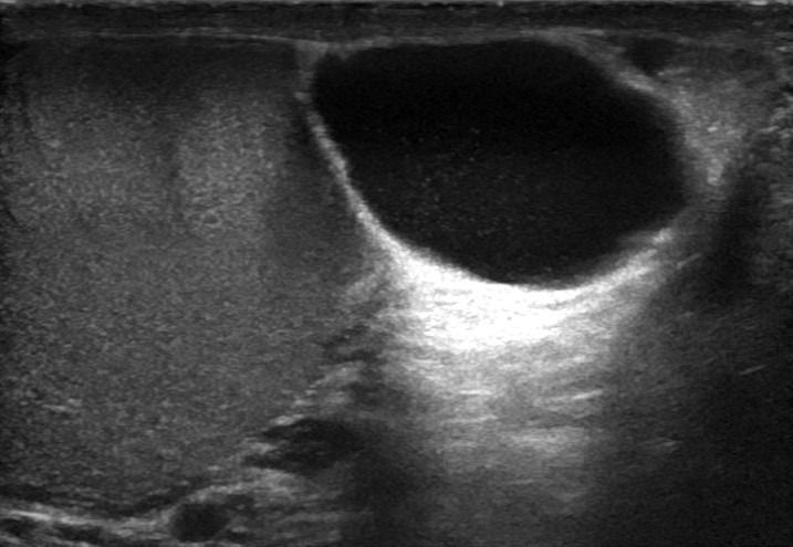
Ultrasound of the Scrotum is particularly effective in diagnosing and evaluating common conditions of the testicles and epididymis, such as:
- Varicocele
- Hydrocele
- Orchitis
- Torsion
- Cysts
- Inflammation
Specialized Ultrasound Techniques
The study with Color Doppler provides critical information about the vascularity of the testicles, detecting torsion or inflammation, such as orchitis.
for detecting varicocele, adding an additional level of accuracy to the diagnosis.
Regular Examinations and Prevention
Regular ultrasound examination of the testicles is an important means of early detection of potential diseases and avoiding further complications.
How is Ultrasound of the Scrotum performed, and what preparation is required?
The Ultrasound of the Scrotum procedure is performed with a special ultrasound probe. During the examination, the patient lies down, and gel is applied to the skin in the area being examined. With the combined reliability of technology and the experience of the specialized Radiologist, the process is painless for the patient. No preparation is required before the examination.


