

")
What Pathologies Can be Visualized with Foot and Ankle Ultrasound?
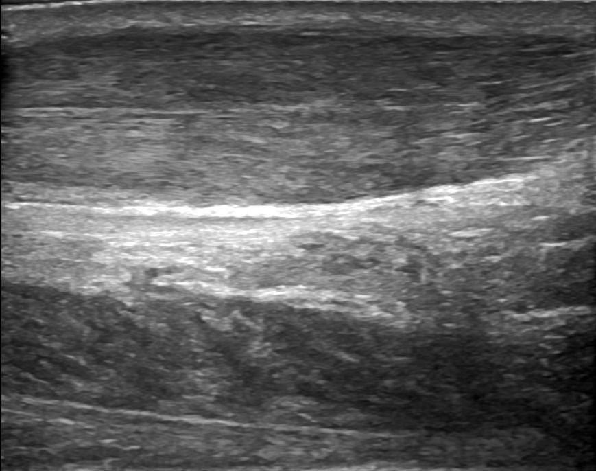
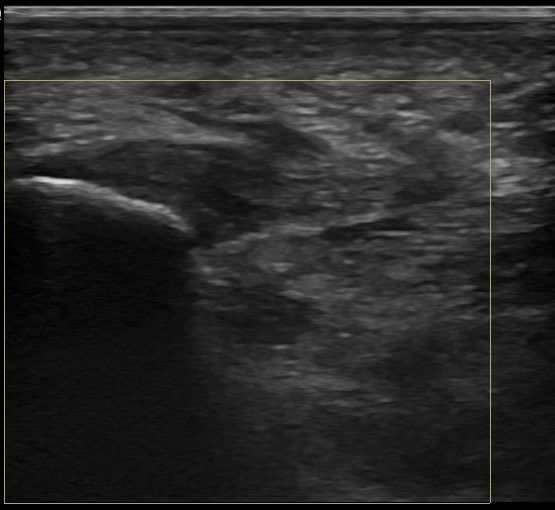
Through Foot and Ankle Ultrasound, a specialized Radiologist can identify issues in the joints, tendons, nerves, and soft tissues of the foot.
Foot Joints
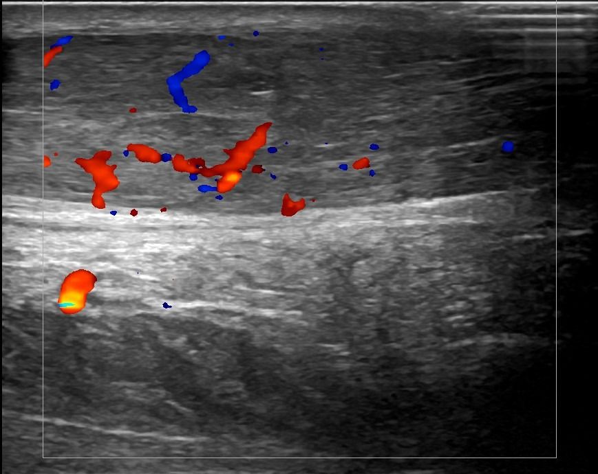
Detection of fluid collection, synovitis, and crystal deposits in the joints of the foot may lead to the diagnosis of rheumatic diseases such as:
- Rheumatoid Arthritis
- Psoriatic Arthritis
- Crystal Arthropathies
- Osteoarthritis
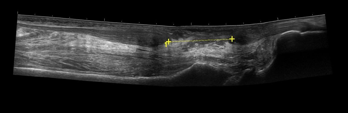
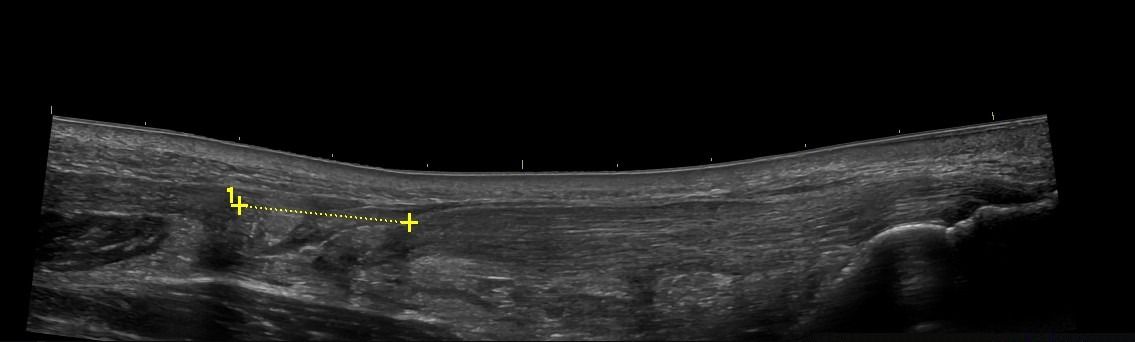
Foot Tendons
Foot Ultrasound allows the diagnosis of tendonitis, tenosynovitis, and rupture in the following tendons:
- Achilles Tendon
- Peroneal Tendons
- Flexor Tendons
- Extensor Tendons
Foot Nerves
- Plantar Fasciitis
- Plantar Fibromatosis
- Morton's Neuroma
- Entrapment syndromes, such as tarsal tunnel syndrome
Soft Tissues
- Lipomas
- Ganglia
- Tumors
Advantages of Foot Ultrasound Compared to Other Imaging Methods
-
Dynamic Testing: The dynamic nature of ultrasound allows for the assessment of the foot during movement. This possibility is particularly useful for evaluating pathologies affecting the range of motion of the foot.
-
Foreign Body Detection: During the ultrasound examination of the soft tissues of the foot, foreign bodies which may not be visible with other imaging methods, such as glass or wood, can be detected.
-
Patient-Friendly Examination: Foot Ultrasound is a painless, safe, and patient-friendly examination that does not involve the use of intravenous drugs, magnetic resonance coordination, or exposure to ionizing radiation.
How is Foot Ultrasound Performed, and What Preparation is Needed?
Foot Ultrasound is a simple and painless procedure that does not require any preparation from the patient. The patient lies on the examination bed, and the radiologist applies gel to the skin in the foot area. The examination is then conducted with a high-frequency ultrasound transducer. During dynamic testing, the patient performs specific movements with their foot as instructed by the radiologist. The total duration of the examination is approximately 30 minutes.


