

")
What can be assessed with the Carotid and Vertebral Artery Triplex?
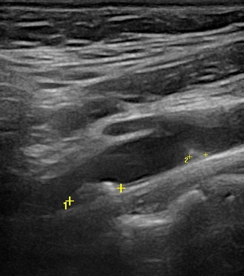
- Carotid Artery Stenosis: Carotid artery stenosis can increase the likelihood of a stroke. The Triplex ultrasound scan helps measure the percentage of stenosis, allowing for the risk assessment.
- Atheromatous Plaques: The quality and stability of atheromatous plaques are critical for predicting the risk of detachment and the occurrence of a stroke. The examination identifies if the plaques are soft and unstable.
What techniques are used in the Carotid and Vertebral Artery Triplex?
For the evaluation of the carotid and vertebral arteries, the following techniques are used:
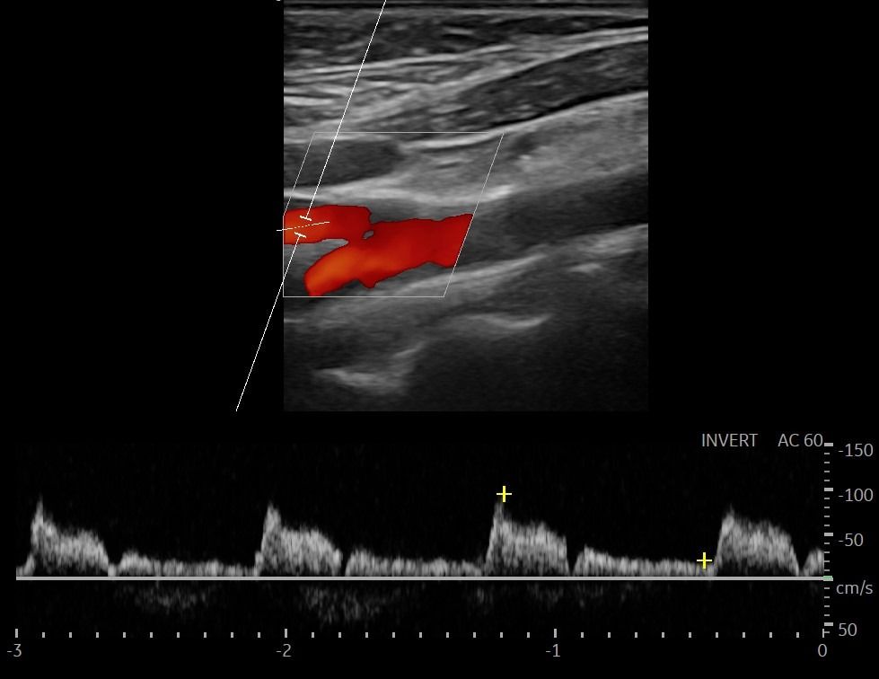
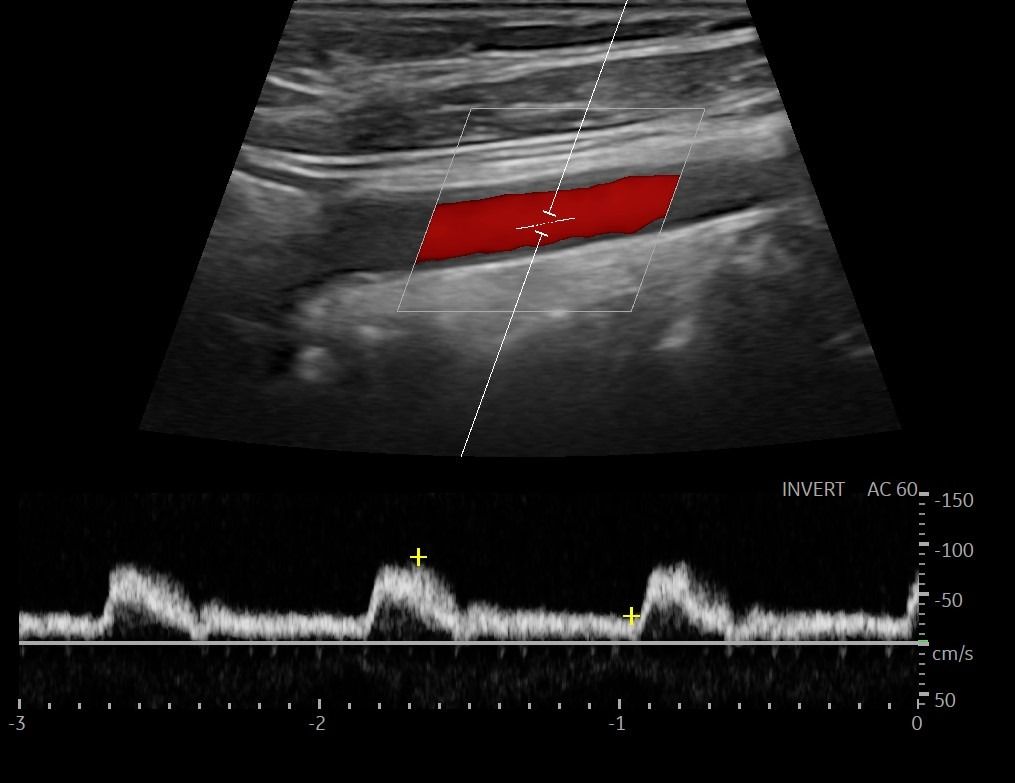
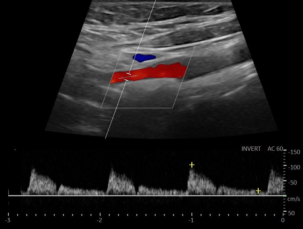
- Color Doppler: It evaluates the speed and direction of blood flow, determining the presence and percentage of arterial stenosis.
- Pulsed Doppler: Provides information about the fluctuations in blood flow, helping identify possible hemodynamic disorders.
- Elastographic Study: Examines the elasticity of the atheromatous plaque, determining if it is soft and unstable, which may influence the likelihood of detachment and lead to ischemic stroke.
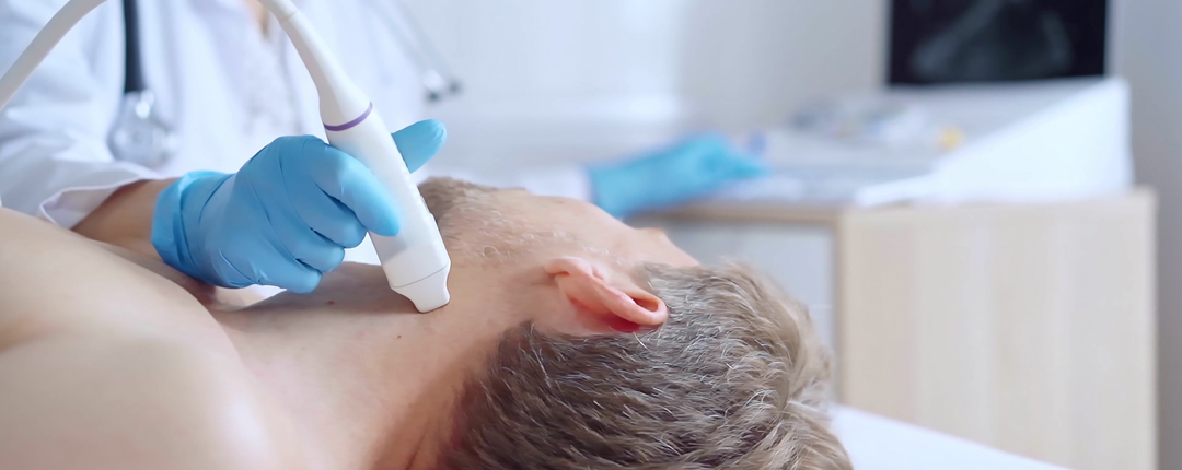
How is the Carotid and Vertebral Artery Triplex performed, and what preparation is needed?
This specific examination requires no preparation. The patient lies in a supine position, and the doctor applies a special ultrasound gel to the examination area. The Carotid and Vertebral Artery Triplex is performed using a high-resolution probe. The procedure is painless and lasts approximately 30 minutes.


